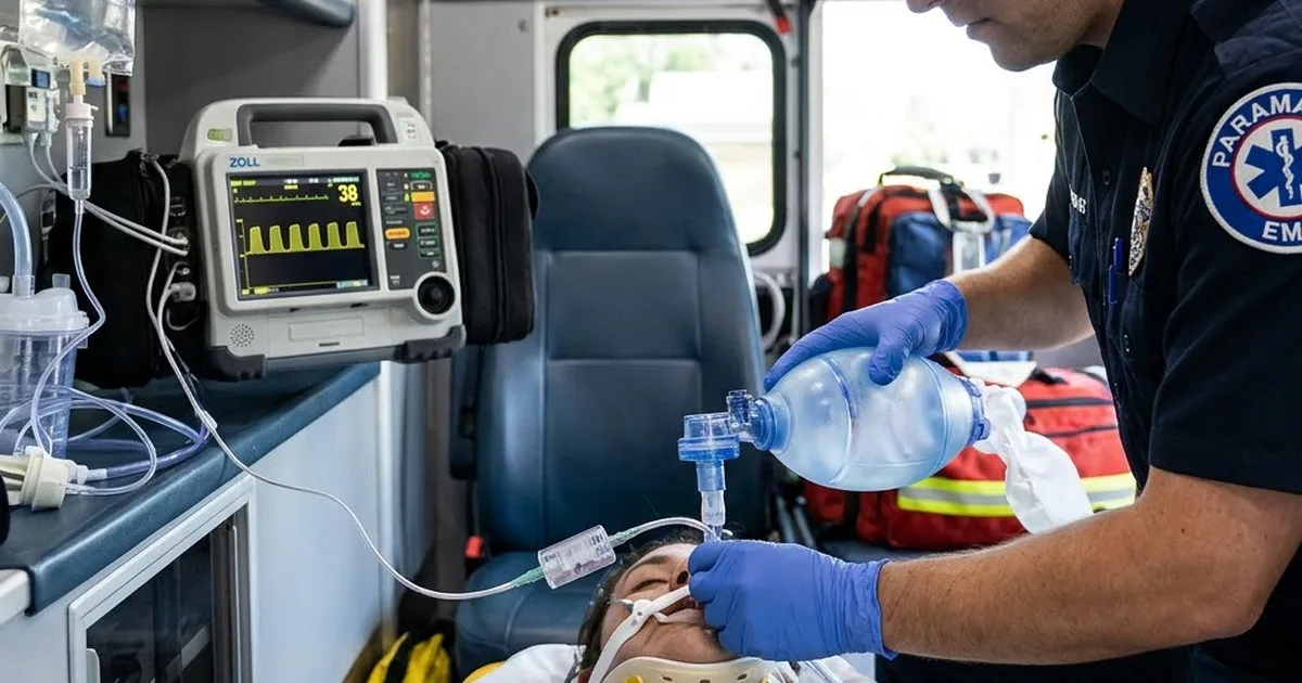
The patient is a 58-year-old man in cardiac arrest on his kitchen floor, compressions running, and your partner has just passed a tube through the cords on the second attempt. You hear breath sounds on both sides. The chest rises with each squeeze of the bag. Then you look at the monitor, and the capnography line is flat. Every instinct in the room wants to blame the sensor.
Do not. A tube with no sustained exhaled carbon dioxide on waveform capnography is in the wrong place until proven otherwise, and the default move is to pull it and ventilate another way.
Why Breath Sounds Cannot Clear a Tube
Prehospital tubes go wrong more often than most crews believe. In a prospective study from a large urban system, emergency physicians checked 108 paramedic-placed tubes on ED arrival and found 25% (27 of 108) misplaced: 18 in the esophagus and 9 above the cords in the hypopharynx [1]. Ten of the 18 patients with esophageal tubes died in the ED [1].
The physical exam does not catch this. The 2022 consensus guideline from the Project for Universal Management of Airways and international airway societies is blunt: misting of the tube, chest rise, lack of abdominal distension, lung and epigastric auscultation, bougie hold-up and chest X-ray should not be used to exclude esophageal intubation [2]. In every unrecognized esophageal intubation the authors reviewed in which auscultation was done, someone reported hearing breath sounds [2].
That is confirmation bias at work. The crew wants the tube to be in, hears what it expects to hear, and explains away the flat trace. The guideline names a second trap, anchoring: deciding that absent carbon dioxide must be due to cardiac arrest or bronchospasm [2]. Both errors start with trusting the exam over the monitor.
What Sustained Exhaled CO2 Means
The 2022 consensus makes detection of sustained exhaled carbon dioxide on waveform capnography the mainstay for excluding esophageal placement [2]. "Sustained" is the key word. Carbon dioxide can appear briefly from an esophageal tube after mask ventilation pushed gas into the stomach, after carbonated drinks or antacids, or after bystander rescue breaths [2]. That gastric carbon dioxide does not hold up breath after breath. A tracheal tube gives a repeating waveform that stays.
The guideline sets a floor. A peak below 7.5 mmHg (1 kPa) is highly unlikely after tracheal intubation in a patient with a pulse [2]. In cardiac arrest, compressions lower the number, but a reading below 7.5 mmHg with good compressions means either a misplaced tube or a very poor outlook [2]. The first move is to check for a pulse and, if there is none, start high-quality compressions. If the peak stays under 7.5 mmHg, the criteria are not met [2]. Arrest does not explain a flat trace [2].
When the criteria are not met, the default action is to remove the tube and ventilate with a bag-valve mask or a supraglottic airway [2]. Leave the tube only if pulling it is clearly more dangerous. Even then, actively exclude esophageal placement at once. The guideline recommends repeat laryngoscopy first, backed by a second valid check such as an esophageal detector device, ultrasound or bronchoscopy [2]. Get a second clinician to do the repeat look if you can: in the cases the authors reviewed, repeat laryngoscopy by the person who placed the tube never detected an esophageal intubation, while a different operator often did [2]. If sustained carbon dioxide does not come back quickly, pull the tube [2]. Breath sounds are not on the list of valid checks.
Colorimetric detectors give one color change at one moment. The consensus calls them less accurate and does not advocate them where electronic monitoring is possible [2]. Continuous waveform capnography is the recommended tool [2].
Confirm Once, Then Watch the Whole Transport
Placement is one moment. The airway has to stay correct for the whole call, and that takes a monitor that never stops watching.
The strongest prehospital data come from a prospective observational study of 153 out-of-hospital intubations arriving at a regional Level I trauma center [3]. Among patients whose crews used continuous end-tidal carbon dioxide monitoring, the unrecognized misplaced intubation rate was zero. Among patients without it, the rate was 23.3% (95% CI 13.4% to 36.0%) [3]. The study was not randomized, and agencies chose their own monitoring, so treat the size of the gap with caution.
The 2022 NAEMSP position statements turn this into a standard. Initial and ongoing confirmation of advanced airway placement must be performed with waveform capnography [4]. After drug-assisted airway management, placement must be confirmed and continually monitored with waveform capnography [5]. For supraglottic airways, confirmation of initial and continuous placement with waveform capnography is strongly encouraged as best practice [6]. In cardiac arrest, where it is available, NAEMSP recommends capnography to confirm and monitor the airway, guide compressions and ventilation, identify return of spontaneous circulation, and help decide when to stop resuscitation [7].
Treat every move as a re-check point: the log roll onto a scoop, the lift to the stretcher, the stair chair, the load into the ambulance or aircraft, the move to the ED bed. A sudden loss of the waveform is a displaced or obstructed tube until you prove otherwise. Check the patient and the tube first and the equipment second. Our airway management review covers the placement side; this post is about keeping the tube where you put it.
What a Medical Director Should Audit
First-pass success measures technique. It says nothing about where the tube was when the crew walked through the ED doors.
The 2022 NAEMSP airway quality management statement asks every agency to measure and report process, outcome and balancing measures [8]. It calls for a non-punitive, educational culture, and for hospitals to send outcome information back to the agency and the crews [8]. For drug-assisted airway programs, NAEMSP asks that quality review include continuous physiologic recordings, capnography among them, wherever they are available [5].
Here is a practical audit set. It is my recommendation, built on those statements, not a published national measure:
- A waveform is present in the monitor file within the first minute after placement.
- A waveform is present at handoff, with the receiving team able to see it.
- The full monitor file is attached to the patient care report for every advanced airway.
- Every loss of waveform during the call has a documented cause and action.
- Every tube left in place without sustained carbon dioxide is reviewed by the medical director.
A narrative that says "placement confirmed by ETCO2" is self-report. The uploaded trace is evidence. Build the review on the trace. If your quality improvement program cannot pull every airway case missing a monitor file in one review cycle, fix that before you trust any airway number it produces.
Key Takeaways
- In one urban system, 25% of paramedic-placed tubes were found misplaced on ED arrival, unrecognized by the transporting crew [1].
- Sustained exhaled carbon dioxide on waveform capnography is the mainstay; a peak below 7.5 mmHg with good compressions does not meet the criteria, even in arrest [2].
- When sustained carbon dioxide is absent, pull the tube and ventilate by bag-valve mask or supraglottic airway unless removal is clearly more dangerous [2].
- Audit the uploaded trace at placement and at handoff, not the narrative. Continuous monitoring was associated with zero unrecognized misplacements in one regional system [3].
FAQ
Is a colorimetric CO2 detector enough to confirm tube placement?
Not where waveform capnography is available. The 2022 consensus guideline calls colorimetric detection less accurate and does not advocate it when continuous electronic monitoring is possible [2]. A color change is also a single moment and cannot warn you when the tube moves during transport.
What ETCO2 reading confirms the tube during cardiac arrest?
No single number confirms it. The 2022 consensus states that a peak below 7.5 mmHg (1 kPa) during high-quality compressions does not meet the criteria for sustained exhaled carbon dioxide, and that cardiac arrest does not explain a flat trace [2]. Look for a repeating waveform that persists breath after breath.
Do supraglottic airways need waveform capnography?
Yes. NAEMSP strongly encourages waveform capnography to confirm initial and continuous supraglottic airway placement [6]. A supraglottic airway can shift during movement just like a tube.
Dr. Chet's Take
I have practiced emergency medicine for more than 25 years, and I serve as medical director for a HEMS and critical care ground transport service. From that seat, this article gets the hard part right. The argument holds. A tube is a hypothesis, and the waveform is the test. What I would add is where the capnography line belongs: on the bag, connected and reading, before the first attempt. When the sensor goes on after the tube, the crew has already decided where the tube is, and the monitor becomes a formality. When it is on from the first breath, the first answer comes from the trace. The same logic applies to the monitor itself. Check, at the start of every shift, that capnography is set to record and that the file uploads. A trace you cannot retrieve protects nobody. And one more point the body leaves out: make the call out loud. "Waveform present, every breath." Your partner hears it, the record hears it, and nobody has to guess later.
That being said, the default to pull the tube is only as safe as the plan behind it. In a falling-saturation airway with a difficult view, nobody wants to remove the only tube they got. The honest answer is that the hesitation is real, and it is exactly the omission bias the guideline warns about. The fix is not courage at the moment of crisis. It is preparation before the first look: a sized supraglottic airway opened on the drape, a bag-valve mask with a good seal ready, and an agreement between partners on who says "no trace, tube out." Without that plan, crews leave doubtful tubes in place. With it, pulling the tube becomes a routine step instead of a defeat.
If you are running this call, put the capnography line on the bag before you pick up the laryngoscope. Open the backup airway before the first attempt, not after the second. Say what you see on the trace after every move, and say it again at the bedside when you hand off, with the receiving team looking at the same screen. If you are the medical director, pull ten random advanced airway cases this month. Open the monitor file for each one and look for the waveform at placement and at handoff. Count how many files are missing, not just how many traces look good, because a missing file is the finding. Share what you find with the crews, the good cases included. The trace decides where the tube is.
— Chester Shermer, MD, FACEP | Emergency Medicine, 25+ Years Clinical Experience | State Surgeon
Continue Your Training
If you're an EMT, paramedic, flight medic, or CCTP (or any clinician treating patients daily) trying to understand how AI will actually impact your clinical practice — not just the hype — I put together a free practical guide. You can download the AI in EM Survival Guide here.
Browse All Courses at Global MedOps Command
Relevant Reading on Global MedOps Command:
How to Avoid Becoming an AI Casualty — Dr. Shermer's guide to navigating AI tools in clinical and operational settings without compromising judgment or patient outcomes.
Emergency Department Efficiency Playbook — Practical systems for throughput, triage optimization, and operational efficiency.
Read more from Dr. Shermer on Medium
Connect with Dr. Shermer: LinkedIn — Chester "Chet" Shermer, MD, FACEP.
References
- Katz SH, Falk JL. Misplaced endotracheal tubes by paramedics in an urban emergency medical services system. Ann Emerg Med. 2001;37(1):32-37. PubMed 11145768
- Chrimes N, Higgs A, Hagberg CA, et al. Preventing unrecognised oesophageal intubation: a consensus guideline from the Project for Universal Management of Airways and international airway societies. Anaesthesia. 2022;77(12):1395-1415. PubMed 35977431
- Silvestri S, Ralls GA, Krauss B, et al. The effectiveness of out-of-hospital use of continuous end-tidal carbon dioxide monitoring on the rate of unrecognized misplaced intubation within a regional emergency medical services system. Ann Emerg Med. 2005;45(5):497-503. PubMed 15855946
- Davis DP, Bosson N, Guyette FX, et al. Optimizing physiology during prehospital airway management: an NAEMSP position statement and resource document. Prehosp Emerg Care. 2022;26(sup1):72-79. PubMed 35001819
- Jarvis JL, Lyng JW, Miller BL, et al. Prehospital drug assisted airway management: an NAEMSP position statement and resource document. Prehosp Emerg Care. 2022;26(sup1):42-53. PubMed 35001829
- Lyng JW, Baldino KT, Braude D, et al. Prehospital supraglottic airways: an NAEMSP position statement and resource document. Prehosp Emerg Care. 2022;26(sup1):32-41. PubMed 35001830
- Carlson JN, Colella MR, Daya MR, et al. Prehospital cardiac arrest airway management: an NAEMSP position statement and resource document. Prehosp Emerg Care. 2022;26(sup1):54-63. PubMed 35001831
- Vithalani V, Sondheim S, Cornelius A, et al. Quality management of prehospital airway programs: an NAEMSP position statement and resource document. Prehosp Emerg Care. 2022;26(sup1):14-22. PubMed 35001828
Related simulation training: EM-Sim · Books by Dr. Shermer