Why Pediatric Assessment Is Different
Children are not small adults. Their physiology, disease presentations, and compensatory mechanisms differ fundamentally from adult patients — and these differences directly affect how you assess and manage them in the prehospital environment.
The most important difference: children compensate extraordinarily well until they don't. A child can maintain near-normal blood pressure through massive fluid loss by dramatically increasing heart rate and peripheral vascular resistance. By the time hypotension appears, the child is in decompensated shock — a late and ominous sign.
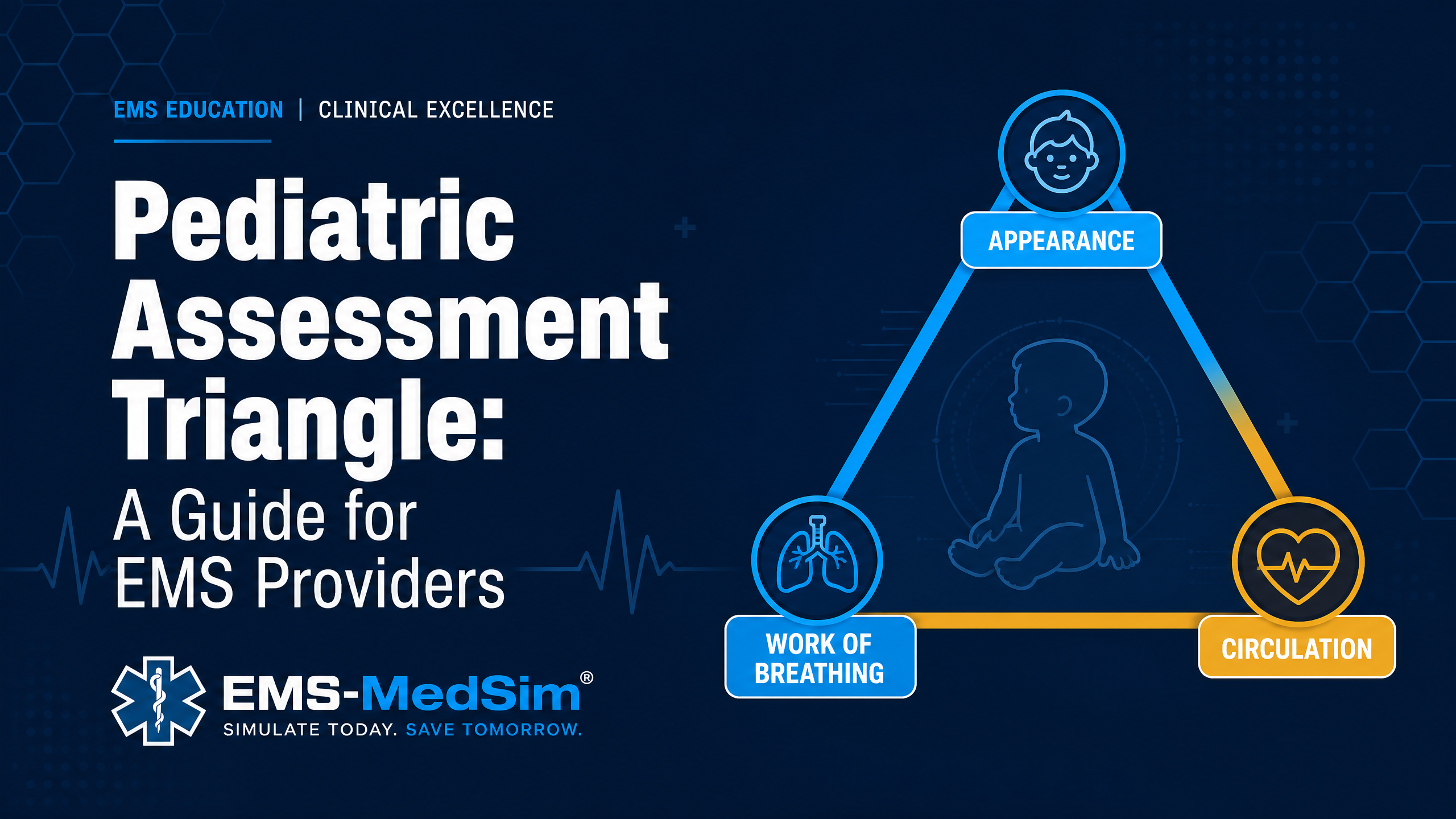
This is why the Pediatric Assessment Triangle (PAT) exists: to identify the sick child before decompensation, using visual cues you can gather in the first 30 seconds without touching the patient.
The Three Components of the PAT
The PAT assesses three domains simultaneously from across the room:
1. Appearance (TICLS)
Appearance reflects the adequacy of cerebral perfusion and oxygenation. Use the TICLS mnemonic:
- Tone: Is the child moving normally? Limp or hypotonic?
- Interactiveness: Is the child engaged with the environment? Responding to you?
- Consolability: Can the child be comforted by a caregiver? Inconsolable crying is abnormal.
- Look/Gaze: Is the child making eye contact? Glazed or vacant stare?
- Speech/Cry: Is the cry strong and appropriate? Weak, high-pitched, or absent?
A child with abnormal appearance has a problem with brain perfusion, oxygenation, or both — regardless of what the rest of the assessment shows.
2. Work of Breathing
Work of breathing reflects the child's respiratory effort and the adequacy of gas exchange. Assess visually for:
- Abnormal sounds: Stridor (upper airway), wheezing (lower airway), grunting (alveolar collapse — very concerning in infants)
- Abnormal positioning: Tripod position, sniffing position, refusal to lie flat
- Retractions: Suprasternal, intercostal, subcostal, or sternal retractions indicate significant respiratory distress
- Nasal flaring: Particularly significant in infants
- Head bobbing: In infants, use of accessory neck muscles creates a head-bobbing motion — a sign of severe respiratory distress
3. Circulation to Skin
Circulation to skin reflects peripheral perfusion and cardiac output. Assess visually for:
- Pallor: Peripheral vasoconstriction shunting blood to core organs
- Mottling: Patchy, irregular skin discoloration — indicates poor perfusion
- Cyanosis: Central (lips, mucous membranes) vs. peripheral (hands, feet)
Note: Skin color assessment is affected by ambient lighting and skin tone. Assess mucous membranes (gums, inner lips) for the most reliable color assessment in patients with darker skin tones.
PAT Interpretation: The Six Clinical Patterns
| Appearance | Work of Breathing | Circulation | Interpretation |
|---|---|---|---|
| Normal | Normal | Normal | Stable — low acuity |
| Abnormal | Normal | Normal | CNS/metabolic problem |
| Normal | Abnormal | Normal | Respiratory distress |
| Abnormal | Abnormal | Normal | Respiratory failure |
| Normal | Normal | Abnormal | Compensated shock |
| Abnormal | Normal | Abnormal | Decompensated shock |
| Abnormal | Abnormal | Abnormal | Cardiopulmonary failure |
Any pattern with abnormal appearance is a high-priority patient requiring immediate intervention.
Age-Specific Normal Vital Signs
One of the most common errors in pediatric assessment is applying adult normal ranges to children. Key reference points:
| Age | Normal HR (bpm) | Normal RR (breaths/min) | Systolic BP (mmHg) |
|---|---|---|---|
| Newborn | 100–160 | 30–60 | 60–90 |
| Infant (1–12 mo) | 100–160 | 25–50 | 70–100 |
| Toddler (1–3 yr) | 90–150 | 20–30 | 80–110 |
| Preschool (3–5 yr) | 80–140 | 20–25 | 80–110 |
| School age (6–12 yr) | 70–120 | 15–20 | 90–120 |
| Adolescent (13+ yr) | 60–100 | 12–16 | 100–130 |
Quick formula for minimum acceptable systolic BP in children 1–10 years: 70 + (2 × age in years)
Weight Estimation for Drug Dosing
Accurate weight estimation is critical for pediatric drug dosing. Options:
- Broselow tape: Color-coded length-based weight estimation. Most validated tool for prehospital use.
- Handspan method: Each handspan ≈ 2.5 kg in children under 10 years (less validated)
- Parent estimate: Ask the caregiver — parents are often accurate within 10%
When in doubt, use the Broselow tape. Overdosing pediatric patients with weight-based medications is a preventable harm.
Building Pediatric Confidence Through Simulation
Pediatric emergencies are low-frequency, high-stakes events. Most paramedics manage fewer than 5 critically ill children per year — not enough real-world exposure to maintain proficiency.
Simulation-based training fills this gap. Branching pediatric scenarios — where your assessment and treatment decisions drive the patient's clinical course — build the pattern recognition and decision-making confidence that transfers to real patients.
Practice pediatric assessment with EMS-MedSim's prehospital simulation scenarios. Try a free scenario — no account required.