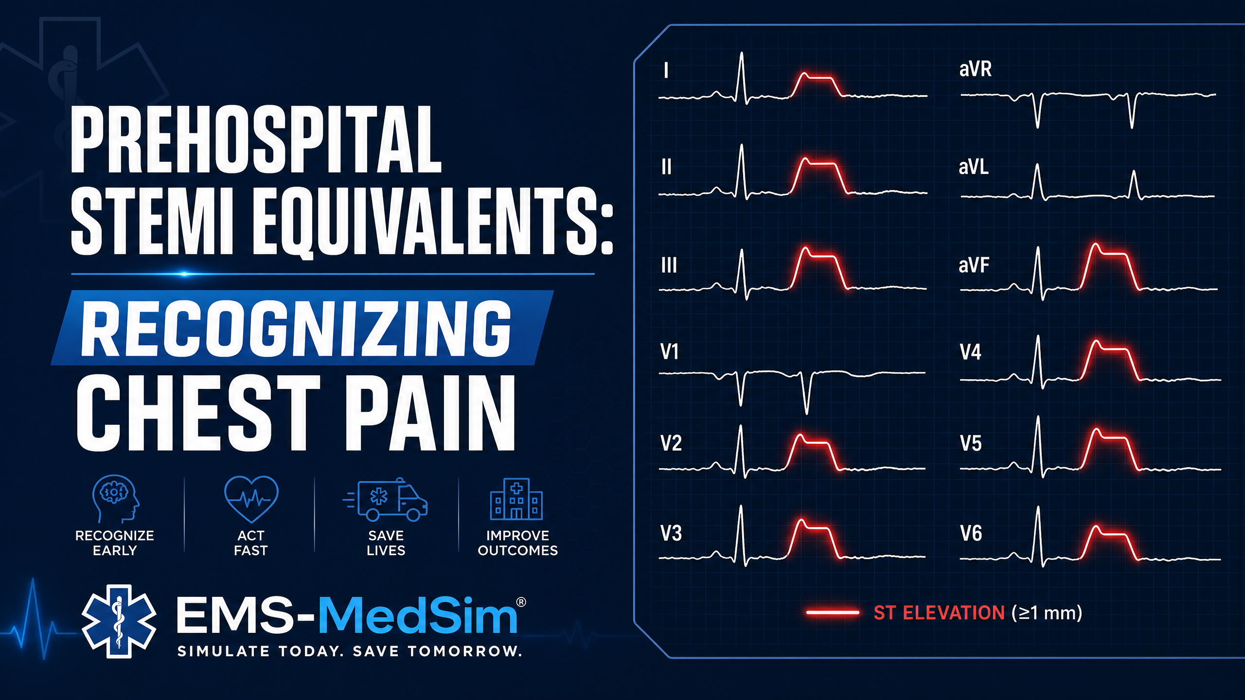
The Problem with "Classic" STEMI Criteria
Every paramedic learns the classic STEMI criteria: ≥1 mm ST elevation in two or more contiguous limb leads, or ≥2 mm in two or more contiguous precordial leads (with lower thresholds in V2–V3 for women and older men). The criteria are easy to teach and easy to put in a protocol. They are also a poor test for the thing that actually matters, which is whether a coronary artery is occluded right now.
The occlusion myocardial infarction (OMI) literature has made the size of the gap hard to ignore. Reviews of the evidence behind the STEMI cutoffs conclude they miss at least one in four acute coronary occlusions and over-triage a similar share to the cath lab (Aslanger, Meyers, Smith, Anatol J Cardiol 2021). In a high-risk acute coronary syndrome cohort of 808 patients, only 41% of the 265 angiographically confirmed occlusions met STEMI criteria on the ECG (Meyers et al., JAHA 2021). The 2022 American College of Cardiology chest pain pathway now lists several of the patterns below as STEMI equivalents that warrant emergent reperfusion (Kontos et al., JACC 2022).
These are the patients who get driven to a non-PCI facility, wait for serial troponins, and lose myocardium that a 12-lead in the ambulance could have saved. Your ability to recognize them changes outcomes. This article covers the patterns, then the field decisions that follow.
Wellens Syndrome: The Warning Before the Infarct
Wellens syndrome represents critical stenosis of the proximal left anterior descending artery (LAD), the "widow maker." It appears on the ECG after an episode of chest pain has resolved, which is exactly why it gets missed: the patient feels better and the medic relaxes.
Two patterns:
- Type A (about 25%): Biphasic T-waves in V2–V3 (positive, then negative deflection)
- Type B (about 75%): Deep, symmetric T-wave inversions in V2–V3
In the original 1982 series of 145 consecutive unstable angina admissions, 18% showed this pattern, and 75% of those managed medically without revascularization went on to an extensive anterior wall infarction within weeks (de Zwaan, Bär, Wellens, Am Heart J 1982).
Why it matters prehospitally: a patient with a Wellens pattern who is currently pain-free is at very high risk of complete LAD occlusion within hours to days. This is not a "watch and wait" presentation, and it is not a patient to talk into a refusal.
Prehospital action: transmit the 12-lead, name the pattern in your prenotification ("biphasic T-waves V2–V3, pain-free now, concerned for Wellens"), and transport to a PCI-capable center even if the patient looks well.
De Winter T-Waves: The STEMI That Doesn't Look Like One
De Winter T-waves represent acute proximal LAD occlusion but appear as upsloping ST depression (≥1 mm) at the J-point in V1–V6 with tall, prominent, symmetric T-waves, not ST elevation. The pattern was described in about 2% of anterior MI presentations and tends to be static rather than evolving into classic STEMI (de Winter et al., NEJM 2008). It is a STEMI equivalent requiring immediate cath lab activation.
Key distinguishing features:
- Upsloping ST depression (not horizontal or downsloping)
- Tall, peaked T-waves following the depression
- Often accompanied by 1–2 mm ST elevation in aVR
- No reciprocal changes in the inferior leads
Hyperacute T-Waves: The Earliest Sign
Before the ST segment rises, the T-wave gets big. Hyperacute T-waves are broad, tall relative to the QRS, and symmetric, and they are often the only finding in the first minutes of an occlusion. They have lacked an objective definition until recently. A five-center derivation and validation study produced one: a T-wave area and symmetry score in two consecutive leads, which was 98.4% specific for acute coronary occlusion in patients without STEMI criteria, and 84% of patients who met the score had a culprit lesion (Meyers et al., JACC Advances 2025).
Field rule: a T-wave that looks too big for its QRS in two neighboring leads, in a patient with an ischemic story, is an occlusion until proven otherwise. Repeat the 12-lead every 5–10 minutes. Hyperacute T-waves evolve, and the second tracing often makes the call for you.
Posterior MI: The Invisible STEMI
The posterior wall of the left ventricle is not directly seen by the standard 12 leads. A posterior MI appears as reciprocal changes in the anterior leads:
- ST depression in V1–V4 (posterior ST elevation seen "backwards")
- Tall, upright R waves in V1–V2 (posterior Q waves seen "backwards")
- Upright T-waves in V1–V2
The specificity of this finding is better than most paramedics were taught. Ischemic ST depression that is maximal in V1–V4, rather than V5–V6, was 97% specific for occlusion MI, and a third of those occlusions had less than 1 mm of depression (Meyers et al., JAHA 2021). Amplitude does not rescue you here. Location does.
Confirmation: posterior leads V7–V9 show ≥0.5 mm ST elevation. If your monitor can acquire them, use them for any patient with anterior ST depression and no clear alternative explanation. If it cannot, transmit what you have and say the words "ST depression maximal in V1 through V4, concerned for posterior occlusion."
Right Ventricular MI: The Preload-Dependent Infarct
Right ventricular (RV) involvement occurs in roughly a third to a half of inferior STEMIs (RCA occlusion). It matters in the ambulance because these patients are preload-dependent: the failing RV needs filling pressure to push blood through the lungs to the left side.
Recognition on the standard 12-lead: ST elevation in II, III, aVF (inferior pattern), elevation in III greater than II, ST elevation in V1, and ST depression in I and aVL. Right-sided lead V4R showing ≥1 mm ST elevation confirms it.
Prehospital implications:
- Hold nitroglycerin in inferior STEMI until RV involvement is excluded. The 2025 ACC/AHA/ACEP/NAEMSP/SCAI guideline carries the same caution (Rao et al., Circulation 2025). A nitrate-induced crash in a preload-dependent patient is a preventable arrest.
- Fluid challenge (250–500 mL isotonic crystalloid) for hypotension, reassessing lung sounds between boluses.
- Treat bradycardia and heart block, which are common with RCA occlusion, before they become the reason for the hypotension.
aVR: The Forgotten Lead
Lead aVR is the one most medics skip. ST elevation in aVR ≥1 mm with diffuse ST depression in six or more other leads points at left main or proximal LAD disease, or at severe three-vessel subendocardial ischemia. This is a high-mortality presentation that needs emergent cardiology evaluation. It is not, by itself, proof of an acute occlusion, which is why some of these patients go to the lab and others go to the operating room. Your job is the same either way: transmit, prenotify, and go to a center that can do both.
Bundle Branch Block and Paced Rhythms: Use the Modified Sgarbossa Criteria
"Can't read a STEMI in LBBB" is a myth that costs lives. The modified Sgarbossa criteria look for ST changes that are concordant with the QRS, or discordant ST elevation that is excessive relative to the depth of the preceding S wave (ST elevation ≥25% of the S wave). In ventricular paced rhythm, the modified criteria were 81% sensitive for occlusion MI against 56% for the original criteria, with 96% specificity (Dodd et al., Ann Emerg Med 2021). A paced or bundle-branch-block ECG in a patient with a good story deserves the same scrutiny as a narrow-complex one.
The Field Decisions That Follow
Pattern recognition is half the job. The other half is what you do in the next ten minutes, and it is where most protocols leave medics without cover.
1. Acquire early and repeat. The 2025 ACS guideline calls for a 12-lead within 10 minutes of first medical contact for any patient with possible ACS, and for EMS transmission or interpretation to drive destination and activation (Rao et al., Circulation 2025). Serial tracings every 5–10 minutes catch the evolving occlusion the first ECG missed.
2. Add leads when the pattern asks for them. Anterior ST depression gets V7–V9. Inferior elevation gets V4R. Both take under a minute and change the treatment.
3. Say the pattern, not just the verdict. "Non-diagnostic ECG" ends the conversation. "Upsloping ST depression V2 through V5 with tall T-waves, I am concerned for de Winter" starts one. Receiving physicians act on specific findings. Transmit the tracing, then describe it in those words.
4. Choose the destination for the occlusion, not the criteria. If your read is a STEMI equivalent and your protocol allows PCI-center destination for "suspected acute coronary occlusion," use it. If your protocol only recognizes criteria-positive STEMI, transport to the PCI center anyway when it is reasonable, prenotify, and document the pattern. Then take the case to your medical director: a protocol that cannot route a de Winter pattern to a cath lab is a protocol that needs a revision, and real cases are what get revisions written.
5. Be careful with nitrates and aggressive with aspirin. Hold nitroglycerin for inferior patterns until RV involvement is excluded, and for any hypotensive patient. Aspirin is still the one drug every one of these patients gets unless there is a true contraindication.
6. Document the read. A documented "concerned for posterior OMI, transmitted 14:32, V7–V9 acquired" protects the patient's timeline and your judgment. A documented "no STEMI" on a tracing that shows STD maximal in V1–V4 protects nobody.
Building a Systematic 12-Lead Approach
Speed and accuracy come from a system, not from memorizing pictures. A reliable prehospital sequence:
- Rate and rhythm — Sinus? Any blocks?
- Axis — Normal, left, right, or extreme?
- Intervals — PR, QRS, QT. Any prolongation? Wide QRS means switch to modified Sgarbossa.
- ST changes — Elevation or depression? Which leads? Where is the depression maximal?
- T-wave morphology — Hyperacute, biphasic, inverted, or symmetric?
- R-wave progression — Tall R in V1–V2 that should not be there?
- Reciprocal changes — Do the ST changes make anatomic sense together?
- aVR — Elevated with diffuse depression?
Simulation-based practice with ECG interpretation scenarios is one of the most effective ways to build this pattern recognition. The more 12-leads you read with immediate feedback, even simulated ones, the faster and more accurate the field read becomes.
Dr. Chet's Take
I have been reading 12-leads at the receiving end of EMS transmissions for a long time, and the single most common failure is not a bad read. It is a good read that never made it out of the ambulance. A medic sees anterior ST depression, feels uneasy, does not have a box on the protocol for "uneasy," and writes "non-diagnostic." Twenty minutes later the ED gets a patient with a normal-looking initial troponin and a circumflex that has been closed since the first tracing. Nobody did anything wrong by the letter of the protocol, and the patient lost an hour of muscle.
The fix is not more patterns to memorize, although the patterns in this article matter and you should know them cold. The fix is language and routing. Teach your crews to say what they see in anatomic words, teach your receiving physicians to listen for those words, and get a line into your protocol that lets a medic take a suspected occlusion to a PCI center without needing the magic millimeters. I have written that line into protocols I sign. It is one sentence. It has never once been abused, and it has paid for itself more than once.
The honest part is this: the criteria we all learned were never designed to find occlusions. They were a convenient threshold that became a quality metric, and the metric took on a life of its own. The occlusion MI literature is correcting that, slowly, and the AI ECG tools coming into the field are going to accelerate it. Until your monitor reads these patterns for you, you are the algorithm. Read every lead, repeat the tracing, add leads when the picture asks, and transmit. Then say the words.
Key Takeaways
- STEMI criteria miss at least a quarter of acute coronary occlusions; in a high-risk cohort only 41% of confirmed occlusions met them.
- Wellens, de Winter, hyperacute T-waves, ST depression maximal in V1–V4, and modified Sgarbossa findings in wide-QRS rhythms are occlusion patterns that need the cath lab now.
- Location beats amplitude: anterior ST depression that is maximal in V1–V4 is 97% specific for occlusion MI, even below 1 mm.
- Inferior STEMI gets V4R before nitroglycerin; a preload-dependent RV infarct can arrest on a nitrate.
- Transmit the tracing and describe the pattern in anatomic words. "Non-diagnostic" ends the conversation; "concerned for posterior occlusion" starts the activation.
- Repeat the 12-lead every 5–10 minutes. The second tracing often makes the call the first one could not.
Frequently Asked Questions
What is a STEMI equivalent?
An ECG pattern that signals acute coronary occlusion without meeting classic ST-elevation criteria. Wellens syndrome, de Winter T-waves, hyperacute T-waves, posterior MI, modified Sgarbossa-positive findings in bundle branch block or paced rhythm, and aVR elevation with diffuse ST depression are the patterns covered in this article.
Why doesn't posterior MI show ST elevation on a standard 12-lead?
The injury pattern projects as a mirror image: ST depression in V1–V4 rather than elevation, with tall R-waves where posterior Q-waves would be. Posterior leads V7–V9 make the elevation visible.
What should EMS do with a suspected STEMI equivalent?
Transmit the ECG, repeat it, add posterior or right-sided leads when the pattern calls for them, describe the specific finding in the prenotification, and transport to a PCI-capable center per local protocol. These occlusions carry the same reperfusion urgency as a criteria-positive STEMI.
Can you diagnose an occlusion in left bundle branch block or a paced rhythm?
Yes. The modified Sgarbossa criteria, which look for concordant ST changes or excessively discordant ST elevation relative to the S wave, identify occlusion MI in both rhythms with high specificity and far better sensitivity than the original criteria.
Sources
- Rao SV, O'Donoghue ML, Ruel M, et al. 2025 ACC/AHA/ACEP/NAEMSP/SCAI guideline for the management of patients with acute coronary syndromes. Circulation. 2025;151(13):e771-e862. https://doi.org/10.1161/CIR.0000000000001309
- Kontos MC, de Lemos JA, Deitelzweig SB, et al. 2022 ACC Expert Consensus Decision Pathway on the evaluation and disposition of acute chest pain in the emergency department. J Am Coll Cardiol. 2022;80(20):1925-1960. https://doi.org/10.1016/j.jacc.2022.08.750
- Meyers HP, Bracey A, Lee D, et al. Ischemic ST-segment depression maximal in V1–V4 (versus V5–V6) of any amplitude is specific for occlusion myocardial infarction (versus nonocclusive ischemia). J Am Heart Assoc. 2021;10(23):e022866. https://doi.org/10.1161/JAHA.121.022866
- Meyers HP, Simančík F, Herman R, et al. Hyperacute T waves are specific for occlusion myocardial infarction, even without diagnostic ST-segment elevation. JACC Adv. 2025;4(10 Pt 2):102120. https://doi.org/10.1016/j.jacadv.2025.102120
- Aslanger EK, Meyers HP, Smith SW. Time for a new paradigm shift in myocardial infarction. Anatol J Cardiol. 2021;25(3):156-162. https://doi.org/10.5152/AnatolJCardiol.2021.89304
- Dodd KW, Zvosec DL, Hart MA, et al. Electrocardiographic diagnosis of acute coronary occlusion myocardial infarction in ventricular paced rhythm using the modified Sgarbossa criteria. Ann Emerg Med. 2021;78(4):517-529. https://doi.org/10.1016/j.annemergmed.2021.03.036
- de Winter RJ, Verouden NJW, Wellens HJJ, Wilde AAM. A new ECG sign of proximal LAD occlusion. N Engl J Med. 2008;359(19):2071-2073. https://doi.org/10.1056/NEJMc0804737
- de Zwaan C, Bär FW, Wellens HJJ. Characteristic electrocardiographic pattern indicating a critical stenosis high in left anterior descending coronary artery in patients admitted because of impending myocardial infarction. Am Heart J. 1982;103(4 Pt 2):730-736. https://doi.org/10.1016/0002-8703(82)90480-x